


2019 Nikon Small World winners show that lots of beauty fits into tiny worlds
Oct 24, 2019
Dunja Đuđić
Dunja Djudjic is a multi-talented artist based in Novi Sad, Serbia. With 15 years of experience as a photographer, she specializes in capturing the beauty of nature, travel, concerts, and fine art. In addition to her photography, Dunja also expresses her creativity through writing, embroidery, and jewelry making.
Share:

Nikon Small World photomicrography competition always amazes us with its fantastic entries. The 2019 edition is no exception. This year’s contest winners have been announced, and they show us just how beautiful and incredible even the tiniest subjects can be.
This is the forty-fifth annual Nikon Small World Photomicrography Contest. This year’s first place was awarded to microscopy technician Teresa Zgoda and recent university graduate Teresa Kugler for their stunning photo of a turtle embryo. It was captured using fluorescence and stereo microscopy, and the final image is a masterful example of image-stitching.

Campbell Hall, New York, USA
Fluorescent turtle embryo
Stereomicroscopy, Fluorescence
5x (Objective Lens Magnification)
“Our goal has always been to show the world how art and science intersect,” said Eric Flem, Communications Manager at Nikon Instruments. “As new imaging and microscopy techniques develop over the years, our winners showcase these technology advances more and more creatively. First place this year is no exception.”
Second place was awarded to Nikon Small World veteran Dr. Igor Siwanowicz for his composite image of three single-cell freshwater protozoans, sometimes called “trumpet animalcules.” He used confocal microscopy to capture the detail of the cilia, tiny hairs used by the animals for feeding and locomotion.

Howard Hughes Medical Institute (HHMI)
Janelia Research Campus
Ashburn, Virginia, USA
Depth-color coded projections of three stentors (single-cell freshwater protozoans)
Confocal
40x (Objective Lens Magnification)
In the third place is Mr. Daniel Smith Paredes, who placed for his image of a developing American alligator embryo. He snapped this photo at around 20 days of development using immunofluorescence and is studying the development and evolution of vertebrate anatomy.

Yale University
Department of Geology and Geophysics
New Haven, Connecticut, USA
Alligator embryo developing nerves and skeleton
Immunofluorescence
10x (Objective Lens Magnification)
This year’s contest received over 2,000 entries from almost 100 countries. Nikon Small World recognized 86 photos out of them, and we bring you the top 20. You saw the first three, and check out the remaining 17 below. Make sure to visit Nikon Small World’s website for honorable mentions and more images from this year’s contest. And if you just can’t get enough, here are links to the winner selections from previous years.

Universität Rostock
Rostock, Mecklenburg Vorpommern, Germany
Male mosquito
Focus Stacking
6.3x (Objective Lens Magnification)

Caleb Foster Photography
Jericho, Vermont, USA
Snowflake
Transmitted Light
4x (Objective Lens Magnification)

Almáchar, Málaga, Spain
Small white hair spider
Reflected Light, Image Stacking
20x (Objective Lens Magnification)

Alicante, Spain
Chinese red carnation stamen
Focus Stacking
3x (Objective Lens Magnification)

Quintin, Cotes-d’Armor, France
Frozen water droplet
Incident Light
8x (Objective Lens Magnification)

Cherkassy, Ukraine
Tulip bud cross section
Reflected Light
1x (Objective Lens Magnification)

Baylor College of Medicine
Optical Imaging & Vital Microscopy Core
Houston, Texas, USA
BPAE cells in telophase stage of mitosis
Confocal with Enhanced Resolution
63x (Objective Lens Magnification)

Kansas State University
Department of Biology
Manhattan, Kansas, USA
A pair of ovaries from an adult Drosophila female stained for F-actin (yellow) and nuclei (green); follicle cells are marked by GFP (magenta)
Confocal
10x (Objective Lens Magnification)

Hounslow, Middlesex, United Kingdom
Mosquito larva
Darkfield, Polarizing Light, Image Stacking
4x (Objective Lens Magnification)

Madrid, Spain
Cuprite (mineral composed of copper oxide)
Focus Stacking
20x (Objective Lens Magnification)

CIRAD – Agricultural Research for Development
Saint Pierre, Réunion
Female Oxyopes dumonti (lynx) spider
Focus Stacking
1x (Objective Lens Magnification)

Marek Miś Photography
Suwalki, Podlaskie, Poland
Pregnant Daphnia magna (small planktonic crustacean)
Modified Darkfield, Polarized Light, Image Stacking
4x (Objective Lens Magnification)

Bucharest, Romania
Housefly compound eye pattern
Focus Stacking, Reflected Light
50x (Objective Lens Magnification)

Eckental, Bavaria, Germany
Vitamin C
Brightfield, Polarized Light
4x (Objective Lens Magnification)

Lotus Gemology
Bangkok, Thailand
Cristobalite crystal suspended in its quartz mineral host
Darkfield
40x (Objective Lens Magnification)

University of Oxford
Weatherall Institute of Molecular Medicine
Oxford, Oxfordshire, United Kingdom
Octopus bimaculoides embryo
Confocal, Image Stitching
5x (Objective Lens Magnification)

University Hospital Essen
Institute for Experimental Immunology & Imaging
Essen, Nordrhein-Westfalen, Germany
Blood vessels of a murine (mouse) heart following myocardial infarction (heart attack)
Tissue Clearing, Light Sheet Fluorescence Microscopy
2x (Objective Lens Magnification)
Filed Under:
Tagged With:

Dunja Đuđić
Dunja Djudjic is a multi-talented artist based in Novi Sad, Serbia. With 15 years of experience as a photographer, she specializes in capturing the beauty of nature, travel, concerts, and fine art. In addition to her photography, Dunja also expresses her creativity through writing, embroidery, and jewelry making.
Related Posts
 Nikon Small World winners show the giant beauty of the microscopic world
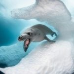 2019 Ocean Art Contest winners show the incredible beauty and diversity of the underwater world
 Bullet Time With Lots And Lots Of Raspberry Pis
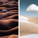 The International Landscape Photographer of the Year winners show all the beauty in the world around us
Join the Discussion
DIYP Comment Policy
Be nice, be on-topic, no personal information or flames.
One response to “2019 Nikon Small World winners show that lots of beauty fits into tiny worlds”
Eugenio