


These are the winning photos from the 2018 Nikon Small World competition, prepare to be amazed
Oct 16, 2018
Dunja Đuđić
Dunja Djudjic is a multi-talented artist based in Novi Sad, Serbia. With 15 years of experience as a photographer, she specializes in capturing the beauty of nature, travel, concerts, and fine art. In addition to her photography, Dunja also expresses her creativity through writing, embroidery, and jewelry making.
Share:

Nikon Small World competition was founded in 1974 to recognize excellence in photography through the microscope. The results of the 44th competition have just been announced, and they will take your breath away.
This year, the contest had nearly 2,500 entries from scientists and artists in 89 countries. The judges have chosen the top 20 images, and we’re bringing you the winning photos here on DIYP.
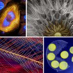
The subjects of the photos vary a lot, which makes the selection even more interesting. In these photos, you can see “everyday stuff” such as human tears, but there are also images of phenomena such as cell division. Still, all these photos have something in common: they show an extreme close-up of their subjects and give us a unique view even on the things we thought we knew.
The judges evaluated the photos on originality, informational content, technical proficiency, and visual impact. First place was awarded to Emirati photographer Yousef Al Habshi, who sees the eyes as the windows to stunning insect artwork and research. The 2018 winning image captures part of the compound eyes and surrounding greenish scales of an Asian Red Palm Weevil. This type of Metapocyrtus subquadrulifer beetle is typically less than 11 mm (0.43 in) in size and is found in the Philippines.

Yousef Al Habshi
Abu Dhabi, United Arab Emirates
Eye of a Metapocyrtus subquadrulifer beetle
Reflected Light
20x (objective lens magnification)
Al Habshi captured the image using a reflected light technique and stacking of hundreds of images: he made a compilation of more than 128 micrographs. According to Al Habshi, “the main challenge was to show the black body against the black background without overexposing the skin and scales.” He was able to strike the perfect balance by controlling the background distance from the subject and using deft lighting and sample positioning.
Second place was awarded to Rogelio Moreno for his colorful photo of a Fern sorus, a clustered structure that produces and contains spores. As for the third place, it was awarded to Saulius Gugis for his adorable spittlebug photo, captured using focus-stacking.

Rogelio Moreno
Panama, Panama
Fern sorus (structures producing and containing spores)
Autofluorescence
10x (objective lens magnification)

Saulius Gugis
Naperville, Illinois, USA
Spittlebug nymph in its bubble house
Focus Stacking
5x (objective lens magnification)
In addition to the top three winners, the Nikon Small World contest has recognized 92 more images from all over the world. We bring you the rest of the top 20 photos below, and you can view the entire gallery, along with Images of Distinction, on the contest’s website. Also, check out the last year’s winners here.
If you’d like to submit the photos of your own, you can do it via this link. The contest is open not only to professionals but to hobbyist photographers as well. And now, let these winning images inspire you to experiment with microphotography yourself.

Can Tunçer
İzmir, Turkey
Peacock feather section
Focus Stacking
5x (objective lens magnification)

Dr. Tessa Montague
Harvard University, Department of Molecular and Cellular Biology
Cambridge, Massachusetts, USA
Parasteatoda tepidariorum (spider embryo) stained for embryo surface (pink), nuclei (blue) and microtubules (green)
Confocal
20x (objective lens magnification)

Hanen Khabou
Vision Institute, Department of Therapeutics
Paris, France
Primate foveola (central region of the retina)
Fluorescence
40x (objective lens magnification)

Norm Barker
Johns Hopkins School of Medicine, Department of Pathology & Art as Applied to Medicine
Baltimore, Maryland, USA
Human tear drop
Darkfield
5x (objective lens magnification)

Pia Scanlon
Government of Western Australia, Department of Primary Industries and Regional Development
South Perth, Western Australia, Australia
Portrait of Sternochetus mangiferae (mango seed weevil)
Stereomicroscopy, Image Stacking
1x (objective lens magnification)

Dr. Haris Antonopoulos
Athens, Greece
Security hologram
Darkfield Epi-illumination
10x (objective lens magnification)

Dr. Csaba Pintér
University of Pannonia, Georgikon Faculty, Department of Plant Protection
Keszthely, Hungary
Stalks with pollen grains
Focus Stacking
3x (objective lens magnification)

Luciano Andres Richino
Punto NEF Photography
Ramos Mejia, Buenos Aires Province, Argentina
Urania ripheus (butterfly) wing scales
Image Stacking
20x (objective lens magnification)

Charles Krebs
Charles Krebs Photography
Issaquah, Washington, USA
Balanus glandula (acorn barnacle)
Autofluorescence
5x (objective lens magnification)

Andrew Moore & Dr. Erika Holzbaur
University of Pennsylvania, Department of Physiology
Philadelphia, Pennsylvania, USA
African green monkey cell (COS-7) stained for actin and microtubules
Stimulated Emission Depletion (STED) Microscopy
100x (objective lens magnification)

Antoine Franck
CIRAD – Agricultural Research for Development
Saint Pierre, Réunion, Reunion Island
Varroa destructor (mite) on the back of Apis mellifera (honeybee)
Focus Stacking
1x (objective lens magnification)

Dr. Amanda D. Phillips Yzaguirre
Salk Institute for Biological Studies
La Jolla, California, USA
Mouse oviduct vasculature
Confocal
10x (objective lens magnification)

Caleb Dawson
The Walter and Eliza Hall Institute of Medical Research, Department of Stem Cells and Cancer
Melbourne, Australia
Breast tissue in lactation: Milk filled spheres (red) surrounded by tiny muscle cells that squeeze out milk (yellow) and immune cells that monitor for infection (blue)
3D Confocal Microscopy
63x (objective lens magnification)

Justin Zoll
Justin Zoll Photography
Ithaca, New York, USA
Amino acid crystals (L-glutamine and beta-alanine)
Polarized Light, Image Tiling
4x (objective lens magnification)

Pierre Anquet
La Tour-du-Crieu, Ariège, France
Vespa velutina (Asian hornet) with venom on its stinger
Reflected Light, Focus Stacking
6.3x (objective lens magnification)

Dr. Nicolás Cuenca & Isabel Ortuño-Lizarán
University of Alicante, Department of Physiology, Genetics and Microbiology
San Vicente del Raspeig, Alicante, Spain
Human retina
Immunocytochemistry and Confocal Microscopy
40x (objective lens magnification)
[All images are courtesy of Nikon Small World and used with permission]

Dunja Đuđić
Dunja Djudjic is a multi-talented artist based in Novi Sad, Serbia. With 15 years of experience as a photographer, she specializes in capturing the beauty of nature, travel, concerts, and fine art. In addition to her photography, Dunja also expresses her creativity through writing, embroidery, and jewelry making.
Related Posts
 Winning photos of 2017 Nikon Small World competition are spectacular
 Winning photos of 2018 Underwater Photographer of the Year are out of this world
 Drone pilot “amazed” to land on deck of Britain’s largest warship without being noticed
Join the Discussion
DIYP Comment Policy
Be nice, be on-topic, no personal information or flames.