


Nikon Small World winners show the giant beauty of the microscopic world
Oct 14, 2022
Dunja Đuđić
Dunja Djudjic is a multi-talented artist based in Novi Sad, Serbia. With 15 years of experience as a photographer, she specializes in capturing the beauty of nature, travel, concerts, and fine art. In addition to her photography, Dunja also expresses her creativity through writing, embroidery, and jewelry making.
Share:

Nikon has announced the winners of its annual Nikon Small World Photomicrography Competition. For almost five decades, Nikon has shared the best microscopic images from this contest, and just as always – they are absolutely marvelous.
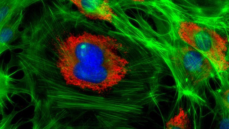
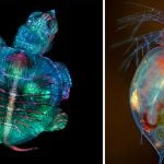
This year’s first-place prize was awarded to Grigorii Timin for his remarkable image of an embryonic hand of a Madagascar giant day gecko. But Nikon also recognizes the best 20 images from the contest, so let’s take a look and enjoy them together.
The Nikon Small World competition was founded in 1974 to recognize excellence in photography through the microscope. In 2011, a sister competition was launched: Nikon Small World in Motion. It was a response to technological advances allowing for recording movies or digital timelapse through the microscope.
For his amazing winning photo, Timin was supervised by Dr. Michel Milinkovitch at the University of Geneva. He utilized high-resolution microscopy and image-stitching to capture this species of Phelsuma grandis day gecko. The final photo is the result of merging hundreds of images together, and preparing the sample was an additional challenge. Timin performed whole-mount fluorescent staining and tissue clearing to capture the entire embryonic hand with a confocal microscope.
“This embryonic hand is about 3 mm (0.12 in) in length, which is a huge sample for high-resolution microscopy,” Timin said. “The scan consists of 300 tiles, each containing about 250 optical sections, resulting in more than two days of acquisition and approximately 200 GB of data.”
The final result gives a glimpse into the hidden beauty and complexity of the gecko, highlighting the nerves in a cyan color and the bones, tendons, ligaments, skin and blood cells in a range of warmer colors.

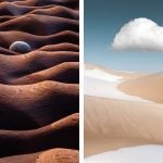
To select the winners, Nikon Small World judges analyzed entries from all over the world. Nearly 1,300 entries from 72 countries, to be more specific. All of the photos were evaluated on originality, informational content, technical proficiency, and visual impact. As I mentioned, the competition has unveiled the top 20 images, which you’ll see if you keep on scrolling. Make sure to check out Nikon Small World’s website for the full gallery of winners and runner-ups.

Breast tissue showing contractile myoepithelial cells wrapped around milk-producing alveoli

Blood vessel networks in the intestine of an adult mouse

Long-bodied cellar/daddy long-legs spider (Pholcus phalangioides)

Slime mold (Lamproderma)

Unburned particles of carbon released when the hydrocarbon chain of candle wax breaks down

Human neurons derived from neural stem cells (NSCs)

Growing tip of a red algae

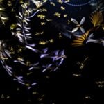
Liquid crystal mixture (smectic Felix 015)

A fly under the chin of a tiger beetle

Moth eggs

Autofluorescence of a single coral polyp (approx. 1 mm)

Agatized dinosaur bone

Differentiated cultured mouse myoblasts with lysosomes (cyan/green), nuclei (yellow), F-actin (magenta)

Cross sections of normal human colon epithelial crypts

Longitudinal section through a white asparagus shoot tip

Tail fin of a zebrafish larva with peripheral nerves (green) and extracellular matrix (violet)

Network of macrophages (white blood cells) of an adult zebrafish intestine

Bacterial biofilm on a human tongue cell

Human cardiomyocytes (heart cells) derived from induced pluripotent stem cells

Dunja Đuđić
Dunja Djudjic is a multi-talented artist based in Novi Sad, Serbia. With 15 years of experience as a photographer, she specializes in capturing the beauty of nature, travel, concerts, and fine art. In addition to her photography, Dunja also expresses her creativity through writing, embroidery, and jewelry making.
Related Posts
 2019 Nikon Small World winners show that lots of beauty fits into tiny worlds
 Nikon Small World in Motion shows us the impressive secrets of microscopic world
 The International Landscape Photographer of the Year winners show all the beauty in the world around us
 Nature inFocus 2022 winners show all the beauty and drama of the natural world
Join the Discussion
DIYP Comment Policy
Be nice, be on-topic, no personal information or flames.