


Winning photos of 2017 Nikon Small World competition are spectacular
Oct 5, 2017
Dunja Đuđić
Dunja Djudjic is a multi-talented artist based in Novi Sad, Serbia. With 15 years of experience as a photographer, she specializes in capturing the beauty of nature, travel, concerts, and fine art. In addition to her photography, Dunja also expresses her creativity through writing, embroidery, and jewelry making.
Share:

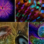
We’ve recently seen the fascinating micro-worlds in the winning videos of Nikon Small World in Motion competition. Now there are also the results of 2017 Nikon Small World photo contest, and they are simply amazing.
Some photos come from scientific labs and show a colorful world of bacteria, algae or cells. But the others show stuff we see every day in a whole new perspective. Have you ever thought mold on a tomato, a credit card hologram or a daddy longlegs’ eyes can look beautiful? Well, the winners of this photo contest show that they can.
The Nikon Small World Competition is open to anyone interested in photography through the microscope. While Nikon is celebrating 100th anniversary this year, the Small World contest has announced winners of the 43rd annual competition. They have received over 2,000 entries from 88 countries around the globe: the United States, Canada, Europe, Australia, Latin America, Asia, and Africa. Winners don’t include only professional photographers, but hobbyists as well.
This year, the first place was awarded to Dr. Bram van den Broek of The Netherlands Cancer Institute (NKI). He took the photo of a skin cell expressing an excessive amount of keratin while he and the associates were researching the dynamics of keratin filaments.

Dr. Bram van den Broek, Andriy Volkov, Dr. Kees Jalink, Dr. Nicole Schwarz & Dr. Reinhard Windoffer
The Netherlands Cancer Institute, BioImaging Facility & Department of Cell Biology
Amsterdam, The Netherlands
Immortalized human skin cells (HaCaT keratinocytes) expressing fluorescently tagged keratin
Confocal
40x (objective lens magnification)
The second place photo was taken by Dr. Havi Sarfaty of Yahud-Monoson from Israel. It captures a subject we see every day, but from a microscopic perspective. It’s the flowering head of otherwise a rather unappealing plant – groundsel (senecio vulgaris). He says he “submitted this photo because of how it represents the unseen complexity of a supposedly simple garden flower.”

Dr. Havi Sarfaty
Eyecare Clinic
Yahud-Monoson, Israel
Senecio vulgaris (a flowering plant) seed head
Stereomicroscopy
2x
The photo that took the third place belongs to Mr. Jean-Marc Bablian of Nantes, France. And no, it’s not Pacman, but a living volvox algae releasing its daughter colonies.

Jean-Marc Babalian
Nantes, France
Living Volvox algae releasing its daughter colonies
Differential Interference Contrast
100x
Take a look at the rest of the top 20 photos from the contest:

Teresa Zgoda
Rochester Institute of Technology
Rochester, New York, USA
Taenia solium (tapeworm) everted scolex
200x

Dean Lerman
Netanya, Israel
Mold on a tomato
Reflected Light, Focus Stacking
3.9x

Dr. David A. Johnston
University of Southampton/University Hospital Southampton, Biomedical Imaging Unit
Southampton, United Kingdom
Lily pollen
Confocal
63x (objective lens magnification)

Dr. Ryo Egawa
Nagoya University, Graduate School of Medicine
Nagoya, Japan
Individually labeled axons in an embryonic chick ciliary ganglion
Differential Interference Contrast
Confocal, Tissue Clearing, Brainbow (labeling technique)
30x (objective lens magnification)

Dr. Michael Perny
University of Bern, Institute for Infectious Diseases
Bern, Switzerland
Newborn rat cochlea with sensory hair cells (green) and spiral ganglion neurons (red)
Confocal
100x

Catarina Moura, Dr. Sumeet Mahajan, Dr. Richard Oreffo & Dr. Rahul Tare
University of Southampton, Institute for Life Sciences
Southampton, United Kingdom
Growing cartilage-like tissue in the lab using bone stem cells (collagen fibers in green and fat deposits in red)
Second Harmonic Generation (SHG) and Coherent Anti-Stokes Raman Scattering (CARS)
20x for collagen; 40x for fat deposits

Dr. Csaba Pintér
University of Pannonia, Georgikon Faculty, Department of Plant Protection
Keszthely, Hungary
Phyllobius roboretanus (weevil)
Stereomicroscopy
80x

Steven Simon
Simon Photography
Grand Prairie, Texas, USA
Plastic fracturing on credit card hologram
10x (objective lens magnification)

Charles Krebs
Charles Krebs Photography
Issaquah, Washington, USA
Opiliones (daddy longlegs) eye
Reflected Light, Image Stacking
20x (objective lens magnification)

Levon Biss
Levon Biss Photography Ltd
Ramsbury, United Kingdom
Exaerete frontalis (orchid cuckoo bee) from the collections of the Oxford University Museum of Natural History
Reflected Light
10x (objective lens magnification)

David Millard
Austin, Texas, USA
Common Mestra butterfly (Mestra amymone) eggs, laid on a leaf of Tragia sp. (Noseburn plant)
Incident Illumination, Image Stacking
7.5x (objective lens magnification)

Dr. Rick Adams
University of Northern Colorado, Department of Biological Sciences
Greeley, Colorado, USA
3rd trimester fetus of Megachiroptera (fruit bat)
Darkfield, Stereomicroscopy
18x

Marek Miś
Marek Miś Photography
Suwalki, Poland
Parus major (titmouse) down feather
Polarized Light, Darkfield
25x

Harald K. Andersen
Steinberg, Norway
Dyed human hair
Darkfield
40x

Christian Gautier
Biosphoto
Le Mans, France
Synapta (sea-cucumber) skin
Polarized Light
100x

Dr. Dylan Burnette
Vanderbilt University School of Medicine, Department of Cell and Developmental Biology
Nashville, Tennessee, USA
Embryonic body wall from a developing Mus musculus (mouse)
100x (objective lens magnification)

Tracy Scott
Ithaca, New York, USA
Aspergillus flavus (fungus) and yeast colony from soil
Transmitted Light
40x
In addition to the first, second and third prize winners, Nikon Small World recognized an additional 85 photos from all the entries they’d received. Other than the first 20 places, there are also honorable mentions and images of distinction. I chose a couple of them I found the most appealing (a tough choice, I must say), and you can see the entire list here.

Emre Can Alagöz
Marmara University, Department of Photography
Istanbul, Turkey
Jumping Spider
Reflected Light
6x

Dr. Regis C. Grailhe, Nasia Antoniou & Dr. Rebecca Matsas
Institut Pasteur Korea, Department of Screening Sciences & Novel Assay Technology
Seongnam-si, South Korea
Neurons derived from a Parkinson patient
Confocal
20x

Dr. Keunyoung Kim
National Center for Microscopy and Imaging Research (NCMIR), Department of Neuroscience
La Jolla, California, USA
Ganglion cells expressing fluorescent proteins in a mouse retina
40x

Dr. Robert Markus
University of Nottingham, School of Life Sciences Super Resolution Microscopy
Nottingham, United Kingdom
Traxacum officinale (dandelion) cross section showing curved stigma with pollen
Confocal, Large Image Stitching
25x

Frank Reiser
Nassau Community College, Department of Biology
Garden City, New York, USA
Human tongue blood vessels injected with lead chromate
Reflected Light, Focus Stacking
100x

Michael Tuchband
University of Colorado Boulder, Department of Physics
Boulder, Colorado, USA
Liquid crystal
Polarized Light
500x

Emilio Carabajal Márquez
Madrid, Spain
Nsutite and Cacoxenite (minerals)
Image Stacking
5x (objective lens magnification)

Dr. Jason Dinelli
Rochester Institute of Technology, School of Photographic Arts and Sciences
Rochester, New York, USA
Water centipede
Differential Interference Contrast, Image Stacking
20x (objective lens magnification)

Dr. Lorenzo Domenis
Istituto Zooprofilattico Sperimentale del Piemonte, Liguria e Valle d’Aosta, Sezione di Aosta/National Reference Centre for Wildlife Diseases
Aosta, Italy
Tracheoles (respiratory tubes) of Apis mellifera (bee)
Brightfield
40x (objective lens magnification)

Frank Fox
Konz, Germany
Rotifers
Darkfield, Transmitted Light
20x (objective lens magnification)

Dr. David A. Johnston
University of Southampton/University Hospital Southampton, Biomedical Imaging Unit
Southampton, United Kingdom
Natural sponge
Confocal
100x

Laurie Knight
Tonbridge, United Kingdom
Simple Eyes of Ectemnius (digger wasp) with condensation
Reflected Light
20x (objective lens magnification)

Henri Koskinen
Helsinki, Finland
Paracetamol (common painkiller) crystals
Polarized Light
20x (objective lens magnification)

Charles Krebs
Charles Krebs Photography
Issaquah, Washington, USA
Mosquito larva, early development
Darkfield
10x (objective lens magnification)

Alexey Marchenko
AlexmarPhoto
Minsk, Belarus
Semiconductor laser diode from a DVD drive
Reflected Light, Image Stacking
3.6x (objective lens magnification)

Dr. Sarah Moghadam & Dr. Ahmad Salehi
VA Palo Alto Health Care System/Stanford University, Department of Psychiatry
Palo Alto, California, USA
Hippocampal region (brain) in an adult mouse
Rapid Golgi staining and automatic stitching
10x (objective lens magnification)

Walter Piorkowski
South Beloit, Illinois, USA
Moth eggs in spider silk
Reflected Light, Image Stacking
16x

Dr. Paul Rigby
The University of Western Australia, Centre for Microscopy, Chracterisation & Analysis
Perth, Australia
Melaleuca sp. (paperbark tree) leaf
Confocal Autofluorescence
100x
As I mentioned, the winners of the contest include both hobbyists and professionals. So if you’re interested in participating, you can apply, too. And even if not, I hope you enjoyed these photos as much as I did.
Filed Under:
Tagged With:

Dunja Đuđić
Dunja Djudjic is a multi-talented artist based in Novi Sad, Serbia. With 15 years of experience as a photographer, she specializes in capturing the beauty of nature, travel, concerts, and fine art. In addition to her photography, Dunja also expresses her creativity through writing, embroidery, and jewelry making.
Related Posts
 These are the winning photos from the 2018 Nikon Small World competition, prepare to be amazed
 These are the cameras that captured the winning shots in this year’s World Press Photo competition
 These are the winning photos of Insight Astronomy Photographer of the Year 2017
 Spectacular Hi-Def Aerial Photos Take You On A Breathtaking Tour Of The World
Join the Discussion
DIYP Comment Policy
Be nice, be on-topic, no personal information or flames.