


The 2015 Wellcome Image Awards Will Spark Your Interest In Science
Mar 10, 2015
Share:

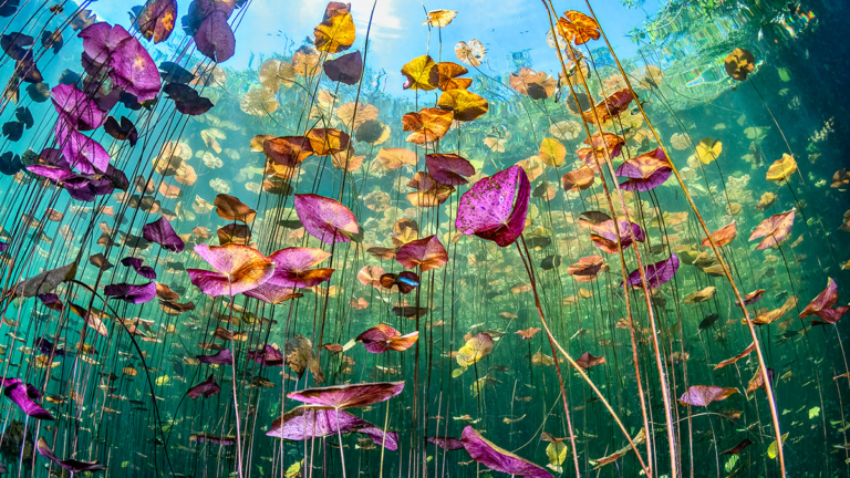
The Wellcome Image Awards recognize science imaging talent and techniques and this year’s winners including some fascinating entries.
Scanning electron micrographs of a boll weevil and a greenfly’s eye, a clinical photograph of an elderly lady’s curved spine and a parasitoid wasp are just a few of the 20 winning images.
The winners were selected from all the images acquired by the Wellcome Images picture library in the past year, and are already accessible along with over 40,000 contemporary biomedical and clinical images.
Unlike other awards, the winning images, along with all content in the Wellcome Images collection, are available under Creative Commons license.

Adam Rutherford, who was one of the judges, spoke about the purpose of the awards:
“The breath-taking riches of the imagery that science generates are so important in telling stories about research and helping us to understand often abstract concepts. It’s not just about imaging the very small either, it’s about understanding life, death, sex and disease: the cornerstones of drama and art. Once again, the Wellcome Image Awards celebrate all of this and more with this year’s incredible range of winning images.”

Catherine Draycott, Head of Wellcome Images and a member of the judging panel added that “This year’s selection of winning images is not only beautiful; they bring to life an incredible array of innovative imaging techniques, and hint at stories and ideas that go beyond the visual.”
Submissions for the 2016 awards are open, but only photos that are accepted into one of Wellcome’s collections can participate.
The 2015 winning gallery will be uploaded next week but until then you can have a look at the 2014 winners.
The overall winner will be announced on March 18 2015.


Arnd Roth, UCL


All images are courtesy Wellcome Images.

Liron Samuels
Liron Samuels is a wildlife and commercial photographer based in Israel. When he isn’t waking up at 4am to take photos of nature, he stays awake until 4am taking photos of the night skies or time lapses. You can see more of his work on his website or follow him on Facebook.


Join the Discussion
DIYP Comment Policy
Be nice, be on-topic, no personal information or flames.