


















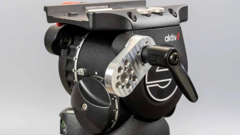

This Microscope Can See Inside Living Antarctic Cells at 0°C
Sep 28, 2026
Soumyadeep Mukherjee
Soumyadeep Mukherjee is an award-winning astrophotographer from India. He has a doctorate degree in Linguistics. His work extends to the sub-genres of nightscape, deep sky, solar, lunar and optical phenomenon photography. He is also a photography educator and has conducted numerous workshops. His works have appeared in over 40 books & magazines including Astronomy, BBC Sky at Night, Sky & Telescope among others, and in various websites including National Geographic, NASA, Forbes. He was the first Indian to win “Astronomy Photographer of the Year” award in a major category.
Share:
A living cell from an Antarctic fish does not behave like one sitting on a microscope slide in a warm laboratory. Its proteins work in extreme cold; its internal machinery has adapted to that environment, and many of its processes operate under conditions that challenge ordinary biological imaging.
Researchers from the British Antarctic Survey and the University of Cambridge have found a solution. They have developed microscopy methods that let them observe living Antarctic fish cells at temperatures near 0°C. The system consists of cold-temperature control with advanced fluorescence imaging. This will allow scientists to examine cellular structures without first moving the cells into warmer conditions.
The researchers had to grow Antarctic fish cells
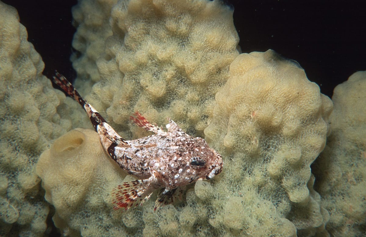
The team worked with the Antarctic spiny plunderfish (Harpagifer antarcticus). This small bottom-dwelling fish lives in shallow waters around the Southern Ocean and sub-Antarctic islands. The surrounding seawater can remain below 0°C, yet the fish continues to function throughout the year.
The researchers developed cell cultures from the plunderfish. According to the British Antarctic Survey, this was the first time scientists had successfully cultured cells from an Antarctic fish for this type of research. The cells grow slowly at temperatures between 0°C and 2°C, which made the process considerably more demanding than maintaining conventional laboratory cell cultures.
The team also cultured cells from the shanny (Lipophrys pholis). This fish lives in shallow waters around the United Kingdom for a comparison with the Antarctic species.
A microscope has to fight the cold too
A live-cell microscope has to maintain focus, collect enough light, and resolve tiny structures while the sample remains stable. Cooling can interfere with each of those requirements.
Materials contract as they cool. This can introduce small movements in the microscope and shift the focus. The cold also changes the behaviour of fluorescent molecules. The researchers use fluorescence to highlight specific structures inside the cells, so the dyes and the optical system have to continue working reliably at low temperatures.
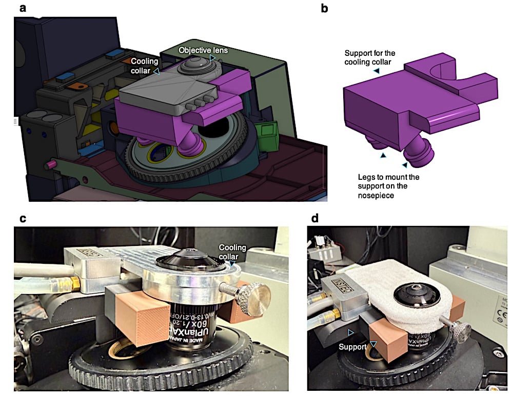
The Cambridge team has spent the project developing ways around these problems. Their work includes advanced fluorescence and super-resolution microscopy adapted for operation at sub-zero temperatures. The research group has also studied the specific limitations that appear when live-cell imaging moves into extreme cold.
The British Antarctic Survey describes the wider project as an effort to study proteins and cellular processes at temperatures of 0°C and below. The researchers want to observe these processes in conditions that resemble the environments where Antarctic organisms evolved.
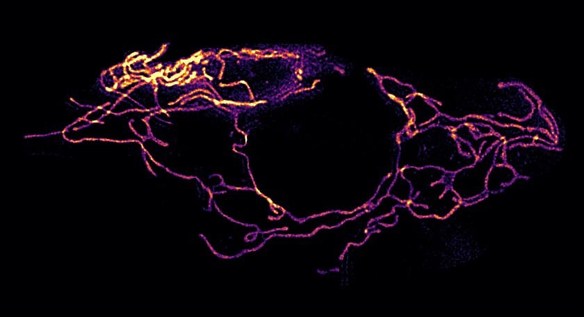
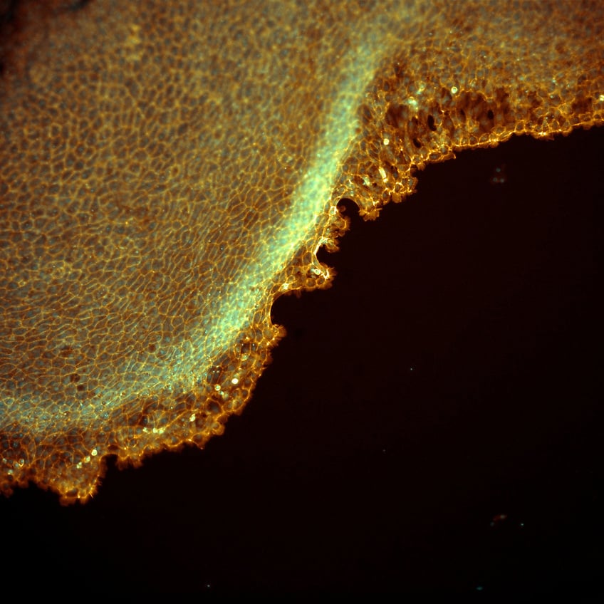
Fluorescent images reveal a very different cell
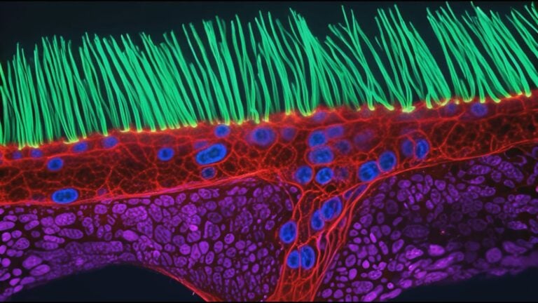
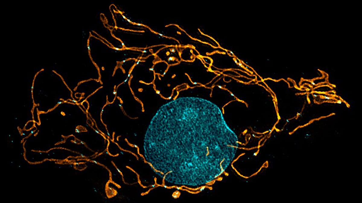
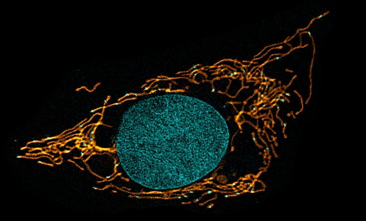
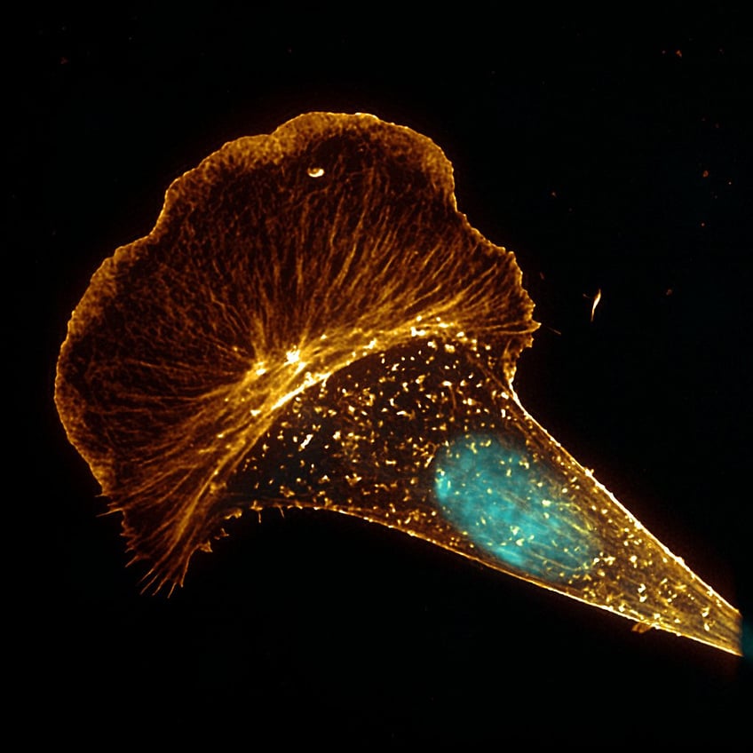
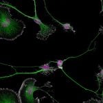
Researchers used fluorescent dyes to label different structures inside the Antarctic fish cells. Under the microscope, these labels made it possible to track organelles and observe how their shape and arrangement changed in cold conditions. The team compared the Antarctic cells with cells from the shanny, a fish found in the much warmer waters around the UK.
One of the clearest differences appeared in the mitochondria. The Antarctic fish cells contained more mitochondria, and many of them had joined together to form larger, connected networks. Mitochondria produce the energy that cells need to function, so having more of them could help the cells maintain their activity when cold temperatures slow many biological processes.
The researchers also found larger lysosomes in the Antarctic cells. These structures break down damaged proteins and other cellular waste. Cold temperatures can make it harder for proteins to fold into their correct shapes, which can leave cells with more damaged or misfolded proteins to remove.
Clear skies!
See more from DIYPhotography.net on Google
Add DIYPhotography.net as a trusted source. This makes
our stories more likely to appear in your Google Search results.
 Add as a preferred source on Google

Soumyadeep Mukherjee
Soumyadeep Mukherjee is an award-winning astrophotographer from India. He has a doctorate degree in Linguistics. His work extends to the sub-genres of nightscape, deep sky, solar, lunar and optical phenomenon photography. He is also a photography educator and has conducted numerous workshops. His works have appeared in over 40 books & magazines including Astronomy, BBC Sky at Night, Sky & Telescope among others, and in various websites including National Geographic, NASA, Forbes. He was the first Indian to win “Astronomy Photographer of the Year” award in a major category.



Join the Discussion
DIYP Comment Policy
Be nice, be on-topic, no personal information or flames.