



















Microscopic Video Revealing a Rare Lung Disease Wins Nikon Small World in Motion 2026
Sep 18, 2026
Dunja Đuđić Kalinin
Dunja Djudjic is a multi-talented artist based in Novi Sad, Serbia. With 15 years of experience as a photographer, she specializes in capturing the beauty of nature, travel, concerts, and fine art. In addition to her photography, Dunja also expresses her creativity through writing, embroidery, and jewelry making.
Share:
The day I anxiously wait for every year has come: Nikon Small World in Motion has announced its 2026 winners. And like every year, the videos are remarkable, with this year’s winner capturing a genetic disorder made visible in real time.
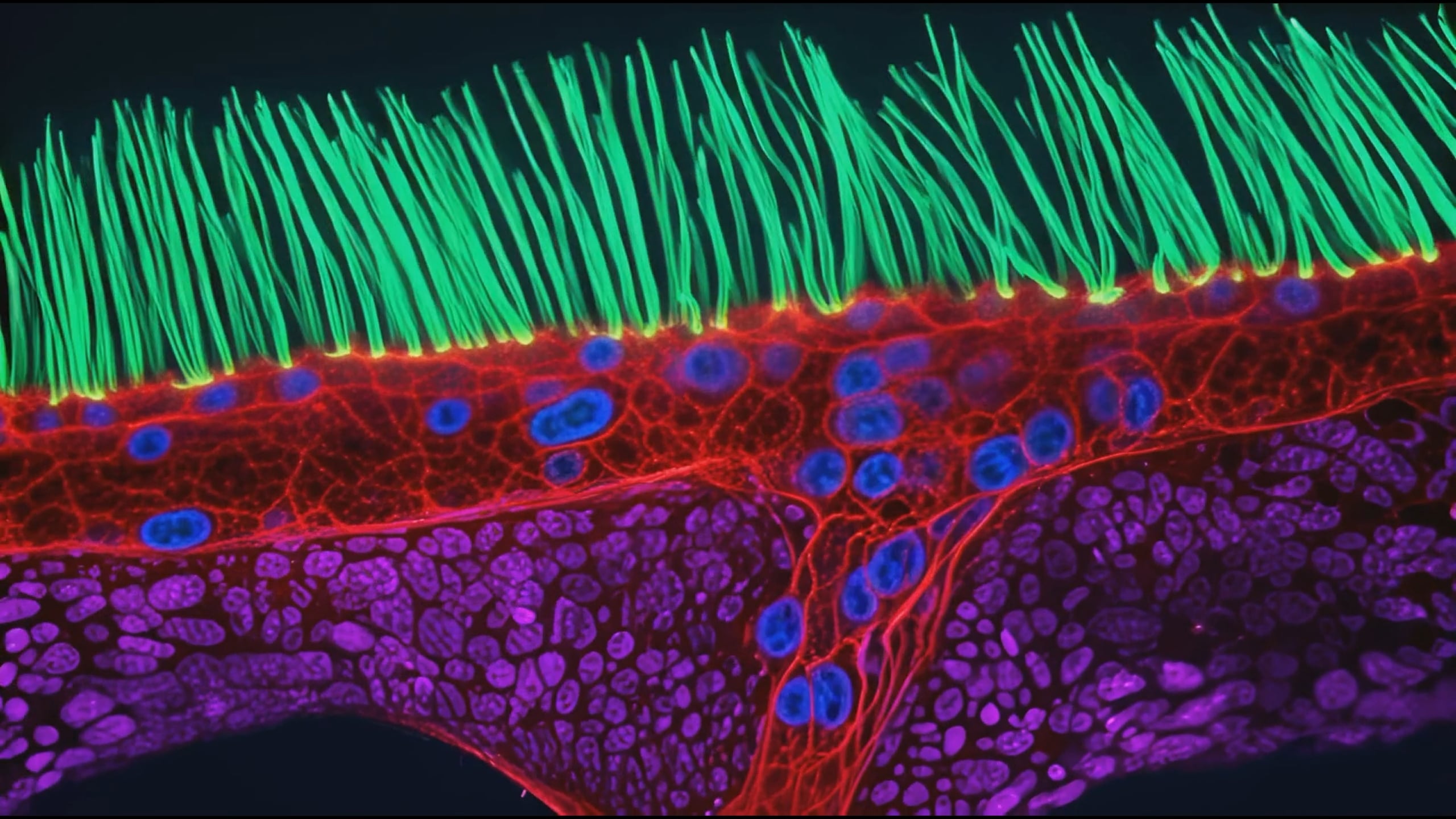
First place goes to Dr. Ning Xu of Tsinghua University in Beijing, China, for a video showing the abnormal beating of airway cilia from a child diagnosed with primary ciliary dyskinesia (PCD). This is a rare condition in which the tiny hair-like structures lining the airways fail to beat in the coordinated waves needed to clear mucus and debris from the lungs. When cilia work the way they should, you barely think about them. When they don’t, the consequences include chronic respiratory infections and serious long-term complications. Ning’s video shows exactly what that dysfunction looks like at 100x magnification, captured using diffractive super-resolution microscopy.
Dr. Ning Xu
Tsinghua University Beijing, Beijing, China
Abnormal beating of airway cilia from a child with Primary Ciliary Dyskinesia (PCD), a genetic disorder affecting respiratory function.
Diffractive Super-Resolution Microscopy
100X (Objective Lens Magnification)
Getting the footage was far from straightforward. The structures are tiny, they move fast, and the sample has to stay alive and behave naturally throughout the process. Many high-resolution microscopy approaches rely on fluorescent labels or intense light, but either can alter or damage living cells. Ning and his collaborators worked around this by controlling the light itself – using multiple light waves and a digital micromirror device to shape illumination precisely, capturing rapid movement at high resolution while keeping the light dose on the sample as low as possible.
“To understand this disease, you need to watch the cilia through dozens of beat cycles, not just a still image,” Ning said.
That point gets at something the competition has championed from the start. Ning put it well:
“For over 50 years, Nikon Small World has repeated one simple message: science is beautiful. And Small World in Motion has a second message that I’m even more passionate about: Life is not a still picture. Life moves.”
He also noted that videos like this one have a communicative power that papers alone don’t. “When people watch cilia beating, they immediately understand that something beautiful and important is happening inside us. I think this competition turns microscopy into a shared language.”
Runner-Ups
Second place went to Nguyen Nam Nhat of Nguyen Nam OME in Chu Se, Gia Lai, Vietnam, for a video captured using differential interference contrast at 40x. It follows a tiny roundworm and a single-celled Dileptus as they navigate their microscopic world.
Nguyen Nam Nhat
Nguyen Nam OME
Chu Se, Gia Lai, Vietnam
A tiny roundworm and a single-celled Dileptus explore their microscopic world
Differential Interference Contrast (DIC)
40X (Objective Lens Magnification)
Third place was awarded to Benedikt Pleyer of Kirchberg, Bavaria, Germany, for a darkfield video of jellyfish larvae suspended in water droplets at 25x. Pleyer picked up two additional honorable mentions this year as well, for a close-up of a tardigrade found on snow and a darkfield shot of a bioluminescent dinoflagellate – a good year by any measure.
Benedikt Pleyer
Kirchberg, Bavaria, Germany
Jellyfish larvae suspended in water droplets
Darkfield
25X (Objective Lens Magnification)
Rounding out the top five: fourth place went to Dr. Andrew Moore of the Howard Hughes Medical Institute’s Janelia Research Campus in Ashburn, Virginia, for a confocal video of synchronized cell division in neighboring cells at 63x. Dr. Patrick Hickey of HYPHA RESEARCH LIMITED in Edinburgh, UK is in the fifth place for confocal footage showing the dynamics of mitochondria and chloroplasts inside Turtle Vine leaf cells at 40x.
Dr. Andrew Moore
Howard Hughes Medical Institute Janelia Research Campus Ashburn, Virginia, USA
Synchronized cell division in neighboring cells
Confocal
63X (Objective Lens Magnification)
Dr. Patrick Hickey
HYPHA RESEARCH LIMITED
EDINBURGH, MIDLOTHIAN, United Kingdom
Dynamics of mitochondria (green) and chloroplasts (red) in Turtle Vine leaf cells (Callisia repens)
Confocal
40X (Objective Lens Magnification)
This year’s competition drew 347 video entries from 40 countries – a remarkable pool judged by a panel that included Dr. Joe Dragavon from the University of Colorado Boulder, Quinten Geldhof of the Museum of Science in Boston, science writer and editor Corey S. Powell, and Dr. Meredith Sagolla from Genentech.
Nikon Small World in Motion was established in 2011 as the video companion to the Nikon Small World Photomicrography Competition. The photo contest was launched in 1974, and the younger video contest is now in its 16th year. There are over 20 honorable mentions in total, and we’ll show you a few more below. But make sure to see the full list at the contest website; it’s definitely worth your time.
[Video courtesy of Nikon Small World]
Charlotte, North Carolina, USA
MSG salt crystals forming in drops of solution
Polarized Light 4X (Objective Lens Magnification)
Vanderbilt University School of Medicine Cell and Developmental Biology Nashville, Tennessee, USA
Sarcomere formation in a heart muscle cell
Confocal
100X (Objective Lens Magnification)
Vanderbilt University Department of Cell and Developmental Biology Nashville, Tennessee, USA
Actin-binding proteins in a polarized epithelial cell after calcium depletion
Confocal 60X (Objective Lens Magnification)
Independent
Edinburgh, Midlothian, United Kingdom
Dung cannon (Pilobolus sp.) growing on fresh manure and then explosively ejecting its sporangium
Reflected Light
10X (Objective Lens Magnification)
CRBM-CNRS
Montpellier, Herault, France
A pregnant tardigrade making contact with another tardigrade, triggering excretion
Brightfield
20X (Objective Lens Magnification)
See more from DIYPhotography.net on Google
Add DIYPhotography.net as a trusted source. This makes
our stories more likely to appear in your Google Search results.
 Add as a preferred source on Google

Dunja Đuđić Kalinin
Dunja Djudjic is a multi-talented artist based in Novi Sad, Serbia. With 15 years of experience as a photographer, she specializes in capturing the beauty of nature, travel, concerts, and fine art. In addition to her photography, Dunja also expresses her creativity through writing, embroidery, and jewelry making.
Related Posts
 Nikon Small World winners show the giant beauty of the microscopic world
 Microscopic Image of a Fat Cell Wins 2026 Endocrine Images Art Competition
 Incredible video of neuron connection wins Nikon Small World in Motion 2023
 Nikon Small World 2025 Winners Reveal the Microscopic Worlds Hidden in Plain Sight
Join the Discussion
DIYP Comment Policy
Be nice, be on-topic, no personal information or flames.